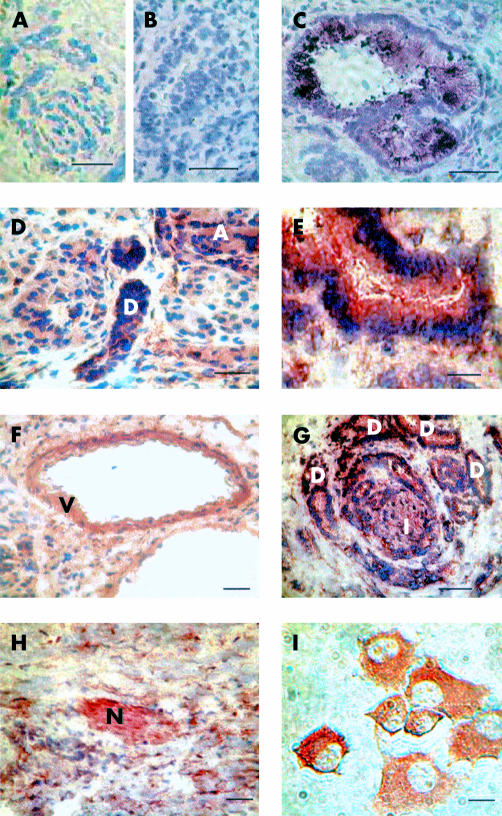
Figure 3.
Immunohistochemistry for calcium sensing receptor (CaR) in normal human pancreas, pancreatic ductal adenocarcinoma, and Capan-1 cells. For human pancreatic samples, 5 μm cryosections were incubated with primary anti-CaR antibody and staining was visualised with diaminobenzidine (DAB) (A, D–I) or the Vector VIP substrate (B, C). Bars represent 200 μm (A, D and F–H) or 50 μm (B,C, E). (A) Control experiment performed in the absence of primary anti-CaR antibody (compare with (D) for example). (B) Preabsorption control (compare with (C)). (C, D, F) Sections from normal human pancreas (“D”=pancreatic duct, “A”=acinus, and “V”=blood vessel). (G, H) Sections from human pancreatic adenocarcinoma (“D”=abundant ductal structures, “I” =islet, and “N”=nerve). Bars represent 200 μm. (E) Enlargement of a portion of the adenocarcinoma section shown in (G) to emphasise heavy staining of ductal structure. (I) Detection of CaR protein in Capan-1 cells. Bar represents 10 μm.

