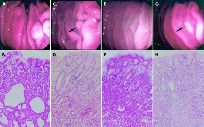
Figure 1.
Endoscopic and microscopic findings in patient No I. Sessile polyps were seen in the normal gastric corpus in February 1999 (A). Biopsies revealed that the polyps consisted of fundic gland hyperplasia with cystic dilatation of glandular ducts—typical morphology of fundic gland polyps (FGPs) (B). Endoscopy one year later showed erythematous mucosa of the corpus and disappearance of FGPs except for one polyp (arrow), the size of which was markedly reduced (C). On histological examination, the remaining polyp showed oedematous changes and neutrophilic infiltration concentrated in the foveolar compartment whereas less inflammatory cells infiltrated the fundic gland compartment (D). Two months after completion of Helicobacter pylori eradication therapy, endoscopy showed reduction of erythematous mucosa and no recurrence of polyps (E). Biopsy demonstrated marked reduction of active inflammation in the remaining polyp (F). Six months after completion of eradication, endoscopy demonstrated enlargement of the remaining polyp (arrow) (G). Biopsy of the polyp revealed hyperplasia of the fundic glands with microcysts, suggesting FGP morphology (H). Endoscopic photographs (top) show the same view in the corpus. (Haematoxylin-eosin staining; original magnifications: lower panels 100×.)

