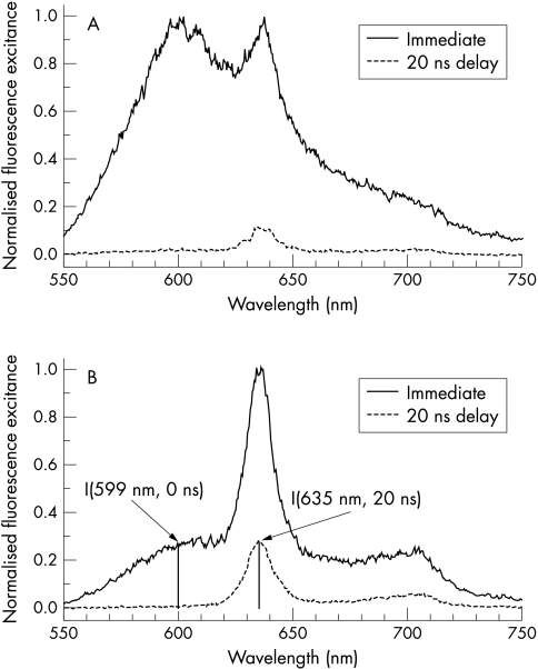
Figure 2.
Fluorescence spectra of non-dysplastic and dysplastic Barrett’s mucosa. Note: the immediate spectra are similar to a certain extent, whereas the delayed spectra of dysplasia and normal mucosa differ conspicuously, in particular by about a factor of 20 in their relative PpIX fluorescence. (A) Typical fluorescence spectra of Barrett’s mucosa. Immediate spectrum: the surrounding mucosa without dysplasia exhibits a broad fluorescence spectrum without any clear spectral signature, and, therefore, the PpIX bands cannot be discriminated from the autofluorescence background. Delayed fluorescence spectrum (20 ns time delay of detection): in the delayed spectrum, a weak PpIX band at 635 nm can be discriminated. (B) Typical fluorescence spectra of dysplasia. Immediate spectrum (0 ns time delay of detection): in the spectrum, well defined fluorescence bands at λ=635 nm and at λ=699 nm appear on top of a broad background, caused by tissue autofluorescence. Delayed fluorescence spectrum (20 ns time delay of detection): because of the difference in the fluorescence decay time of PpIX (τ=16 ns) and of autofluorescence (τ about 2–4 ns), the fluorescence spectrum is virtually free of autofluorescence background and exhibits PpIX fluorescence bands only. For comparison, the prompt spectrum was normalised to the maximum intensity and the corresponding delayed spectrum was corrected for different sampling time and excitation energy.

