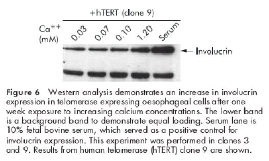
Figure 6.

Western analysis demonstrates an increase in involucrin expression in telomerase expressing oesophageal cells after one week exposure to increasing calcium concentrations. The lower band is a background band to demonstrate equal loading. Serum lane is 10% fetal bovine serum, which served as a positive control for involucrin expression. This experiment was performed in clones 3 and 9. Results from human telomerase (hTERT) clone 9 are shown.
