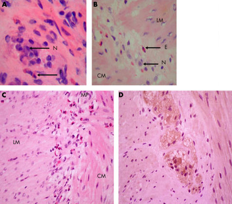
Figure 2.
(A) Haematoxylin/eosin (H/E) stained frozen section of full thickness rectal biopsy from patient No 1 demonstrating an inflammatory infiltrate within the myenteric plexus. N, neurone; E, eosinophil. (B) H/E stained section of a full thickness colonic biopsy from patient No 2 demonstrating eosinophils in the myenteric plexus. LM, longitudinal muscle; CM, circular muscle. (C) H/E stained section of the myenteric plexus and muscularis propria in the colon of patient No 3. Note eosinophils in the myenteric plexus and muscularis. MP, myenteric plexus. (D) Colonic seromuscular biopsy of patient No 2 immunostained for interleukin 5 showing intense positive staining of myenteric neurones.

