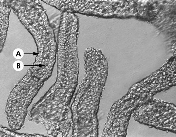
Figure 1 .
Photomicrograph of intact colonic crypts isolated by Ca2+ chelation and visualised using Hoffman modulated optics (×400). Note that the crypts were isolated devoid of non-epithelial cells originating from the lamina propria. The lumenal opening and base of each crypt can be clearly distinguished. Arrows indicate the continuous layer of crypt epithelial cells (A) and the crypt lumen (B).

