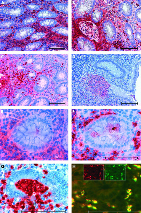
Figure 1 .
Expression of S100A12 in tissues from patients with active Crohn’s disease (CD) or ulcerative colitis (UC). Immunohistochemical staining showed extensive expression of S100A12 in inflamed colonic tissue of patients with active CD (A). S100A12 positive cells surrounded granulomatous lesions in CD (B). Similar local expression of S100A12 was found in UC (C). Numerous S100A12 positive cells assembled in crypt abscesses in UC (D). Staining of serial sections revealed colocalisation of S100A12 positive cells (E) and CD15 positive cells (F). In destructive crypt abscesses, S100A12 positive neutrophils transmigrated through the epithelium into the lumen (G). Immunofluorescence microscopy of double labelling studies with a-S100A12 Texas Red (red) and a-CD15-FITC (green) clearly proved expression of S100A12 by infiltrating CD15 positive granulocytes (H). Double labelled cells appear yellow due to summation of colours. The inserted figures in (H) show emission at a single wavelength for both fluorochromes. Scale bars indicate 100 μm.

