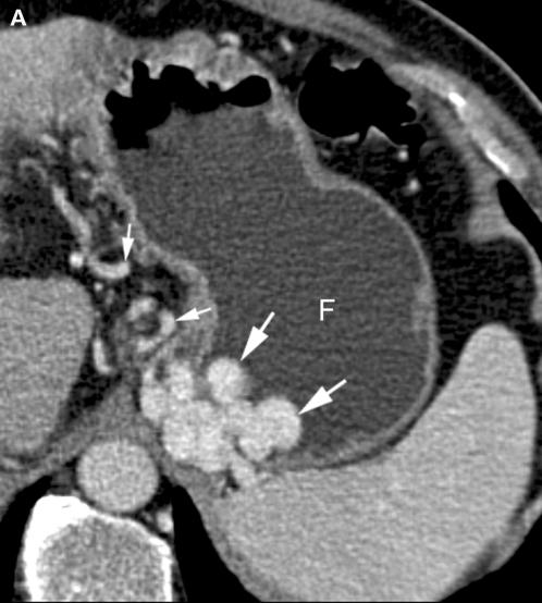
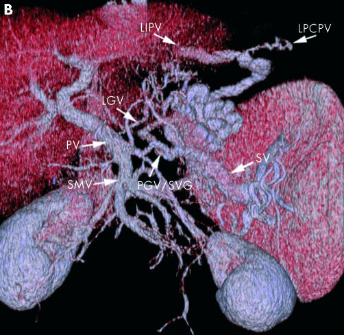
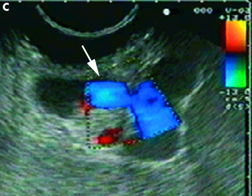
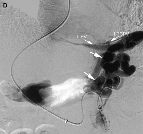
Figure 2 .
Submucosal and perigastric fundal varices in a 68 year old male with alcohol related liver cirrhosis Child-Pugh class A. (A) Transverse source image of multi-detector row CT (MDCT) angiography during the portal-venous phase shows a conglomerate of submucosal varices ≥5 mm in diameter (large white arrows) located on both medial and lateral borders of the water filled gastric fundus (F). Perigastric varices (small white arrows) are also noted. (B) Volume rendering of MDCT angiographic data set during the portal-venous phase demonstrates the submucosal fundal varices with afferent vessels, including the left gastric vein (LGV) and the posterior/short gastric veins (PGV/SGV). The efferent veins drain through the left inferior phrenic vein (LIPV) and the left pericardiacophrenic vein (LPCPV). PV, portal vein; SV, splenic vein; SMV, superior mesenteric vein. (C) Corresponding endoscopic ultrasound image of the same patient confirms the presence of submucosal fundal varices, visible as large hypoechoic vessels (arrow) with continuous colour Doppler flow within the wall. (D) Direct selective digital subtraction venogram of the posterior/short gastric vein after transjugular intrahepatic portosystemic shunt stent placement in the same patient demonstrates a conglomerate of fundal varices (arrows) with the efferent vessels draining through the left inferior phrenic (LIPV) and the left pericardiophrenic veins (LPCPV).


