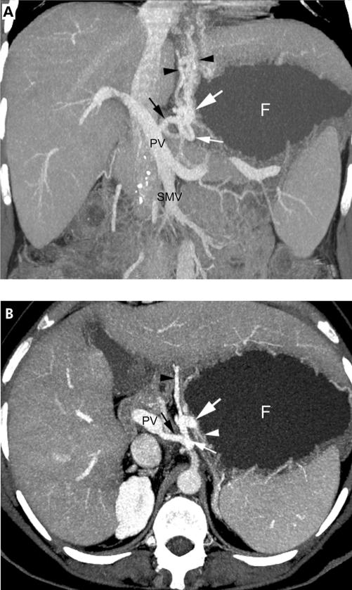
Figure 3 .
A 31 year old female with portal hypertension due to hepatitis C induced liver cirrhosis Child Pugh class A and the presence of submucosal and perigastric fundal varices. (A) Coronal thin slap maximum intensity projection of multi-detector row CT (MDCT) angiography during the portal-venous phase shows a large submucosal varix (≥5 mm in diameter) (large white arrow) located on the medial border of the water filled gastric fundus (F). A perigastric varix (small white arrow) is also noted. The afferent vessel includes the left gastric vein (black arrow) and efferent vessels drain through the oesophageal veins (black arrowheads). PV, portal vein; SMV, superior mesenteric vein. (B) The submucosal location within the gastric wall (white arrowhead) of the varix (large white arrow) is more visible on the transverse thin slap maximum intensity projection.

