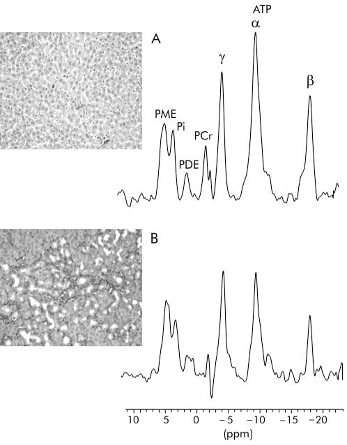
Figure 3.
Representative histological section of liver and corresponding hepatic 31P magnetic resonance spectrum from (A) a control rat and (B) a rat with cholestatic (common bile duct ligation) induced cirrhosis. Van Gieson, ×40 (see methods for description of histological staging). PME, phosphomonoesters; Pi, inorganic phosphate; PDE, phosphodiesters; ATP, adenosine triphosphate. PCr, phosphocreatine resonance arising from abdominal wall muscle.

