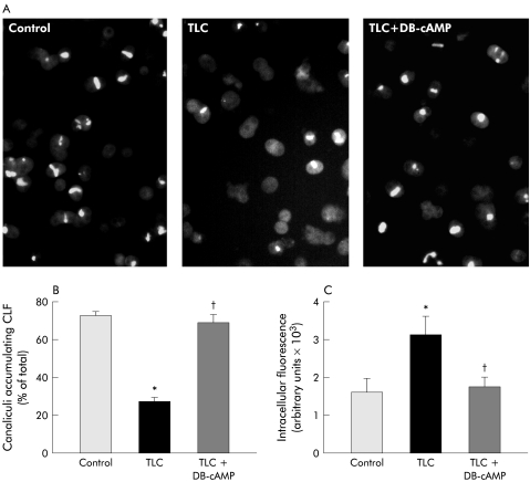
Figure 5.
(A) Representative microphotographs showing accumulation of the fluorescent bile salt analogue cholyl-lysylfluorescein (CLF) in control hepatocyte couplets, and in taurolithocholate (TLC; 2.5 μM, 20 minutes) treated couplets, which were pretreated or not with dibutyryl-cAMP (DB-cAMP; 10 μM, 30 minutes). (B) Quantification of the percentage of total couplets (>50) displaying visible CLF fluorescence in their canalicular vacuoles. (C) Quantification of total pixel intensity of CLF fluorescence in the cellular body of 30 hepatocyte couplets, as a measure of intracellular CLF content. Values are expressed as mean (SEM) obtained from four independent cell preparations. *Significantly different from the control group (p <0.05); †significantly different from the TLC group (p<0.05).

