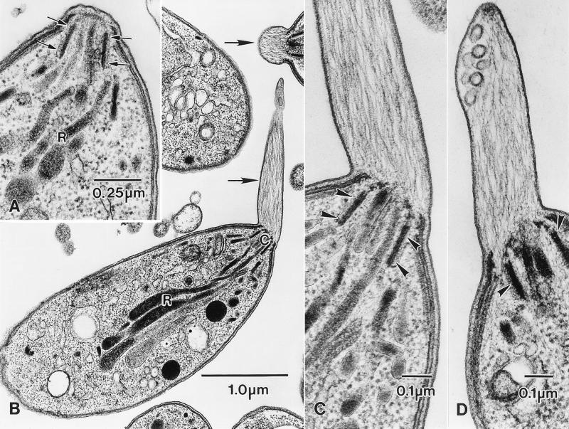
Figure 1.
Jasplakinolide induces the formation of actin-containing apical extensions in isolated T. gondii tachyzoites. (A) High magnification of the apical end of a tachyzoite from an untreated control culture showing the walls of the conoid (arrows) with the necks of the rhoptries (R) closely associated with it. Apart from some fuzzy material associated with the conoid, there is no filamentous material in any part of the apical region. (B) Low-power electron micrograph of isolated tachyzoites treated with 1 μM jasplakinolide for 60 min at 37°C. Filament-containing apical extensions are present in two of the parasites (arrows). C, conoid complex; R, rhoptries. (C and D) Higher-magnification micrographs of the apical end of jasplakinolide-treated tachyzoites showing the membrane-bounded apical extensions, each of which contains many filaments extending from the conoid. Note also the presence of membrane-bounded vesicles and membrane-associated dense material at the anterior end of the protrusions (D). In C and D, arrowheads denote the walls of the conoid.

