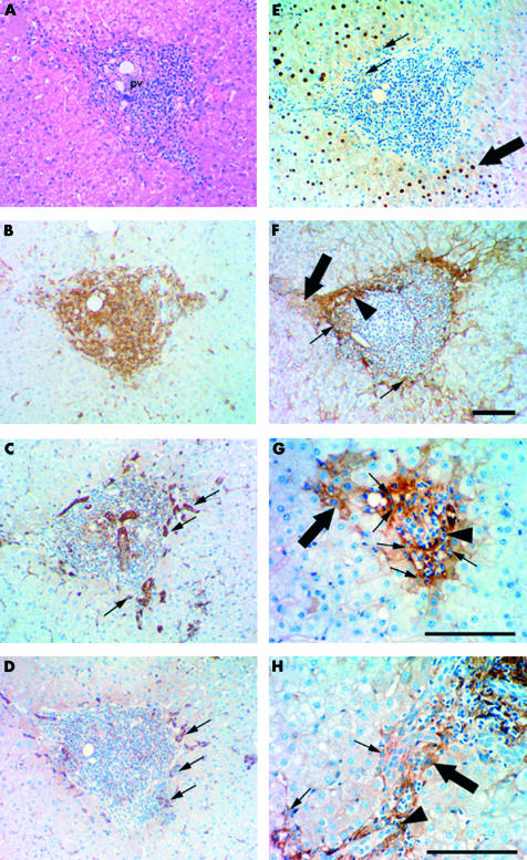
Figure 2.
Immunohistochemical staining illustrating cellular localisation of lymphotoxin β (LT-β) in human hepatitis C virus (HCV) infected liver with bridging fibrosis. (A–F) Serial staining for haematoxylin and eosin (H&E), leucocyte common antigen (LCA) (CD45), π-glutathione S-transferase (GST), M2-pyruvate kinase (M2-PK), proliferating cell nuclear antigen (PCNA), and LT-β suggests oval cells in HCV infected liver express LT-β. (A) H&E staining illustrating periportal cuffing by lymphocytes in chronic hepatitis C. The portal vein is labelled (pv). (B) LCA (CD45) expression illustrates that the mass of small cells clustered around the portal vein are CD45 positive. Oval cells (small arrows) were identified towards the periphery of this region by expression of π-GST (C) and M2-PK (D). (E) The small cells clustered directly around the portal vein are PCNA negative while oval-like cells (small arrows) are weakly PCNA positive and large centrally located hepatocytes (large arrow) are strongly PCNA positive. (F) LT-β expression was restricted to the periphery of the inflammatory cell mass, suggesting that the majority of LCA positive cells are not expressing LT-β. Cell types expressing LT-β include small portal hepatocytes (large arrow), oval cells (small arrows), and inflammatory cells (arrowhead). Bile ducts and large hepatocytes are negative. (G) Periportal cuffing by lymphocytes in chronic hepatitis C, portal fibrosis. This region contains LT-β positive oval cells (small arrows) and small hepatocytes adjacent to the portal vein with membranous and cytoplasmic LT-β staining (large arrow). LT-β expression is observed in some inflammatory cells (arrowhead). (H) Chronic hepatitis C with cirrhosis shows similar LT-β staining patterns, with oval cells (small arrows) and some inflammatory cells (arrowhead) staining positive for LT-β. Hepatocytes adjacent to fibrous septae also express LT-β (large arrow). Magnification bars represent 100 μm.

