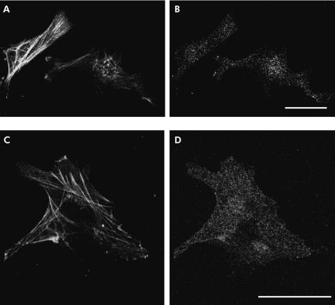
Figure 5.
Fluorescence micrographs showing the immunoreactivity of activin A in pancreatic stellate cells (PSCs). Cells were double stained with an anti-activin A rabbit polyclonal antibody (B, D) and an anti-α smooth muscle actin (α-SMA) mouse monoclonal antibody (A, C). Activin A immunoreactivity was observed in PSCs identified with their α-SMA fine network architecture. Bars 40 µm.

