Figure 1.
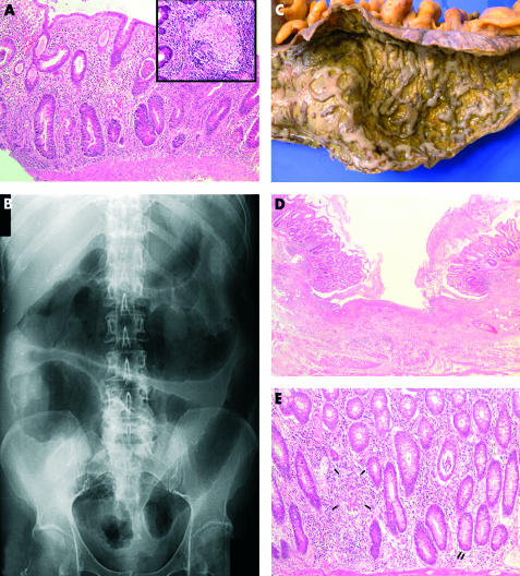
(A) Colonic biopsy showing crypt distortion, patchy transmucosal inflammation, crypt abscesses, and mucin depletion. Inset shows an epithelioid granuloma in the deep mucosa (haematoxylin and eosin; magnification ×100 and ×200 (inset)). (B) Supine plain abdominal x ray with features of toxic megacolon. (C) Macroscopic appearance of the colectomy specimen revealing confluent geographical ulcers covered with pus and separated by small irregular islands of residual mucosa. (D) Microscopy of the colectomy specimen confirming the presence of broad ulceration flanked by inflamed mucosa displaying crypt distortion (haematoxylin and eosin; magnification ×25). (E) Colonic mucosa containing irregular and bifid crypts, occasional epithelioid granulomata (arrows), metaplastic Paneth cells (double arrow), and crypt abscesses (haematoxylin and eosin; magnification ×100).

