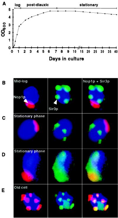
Figure 1.
Stationary-phase cells do not resemble replicatively aging mother cells. (A) Growth curve in YPD at 24°C. (B) Multilabel immunohistochemical study of cells harvested at mid-logarithmic phase. Nop1p (fibrillarin) appears red and marks the nucleolus. Sir3p is stained green and is located outside the nucleolus and is telomeric. DNA is stained blue with DAPI. (C) Typical cells (80–85% of the population) present in 3-week-old stationary-phase cultures. Sir3p is present at telomeres. The nucleolus is crescent shaped and intact. (D) Representative of a subpopulation of cells present in a 3-week-old stationary-phase culture. Sir3p is redistributed throughout the nucleus, including the nucleolus (yellow). Staining patterns shown in B–D were scored in several hundred cells per aliquot per time point per experiment (n = 2 independent experiments). (E) Old cell recovered from a nonstarved culture by sorting (see Materials and Methods). The average bud scar count of the preparation was 15.5 ± 3.3. There is nucleolar fragmentation and redistribution of Sir3p to these fragments.

