
Figure 1.
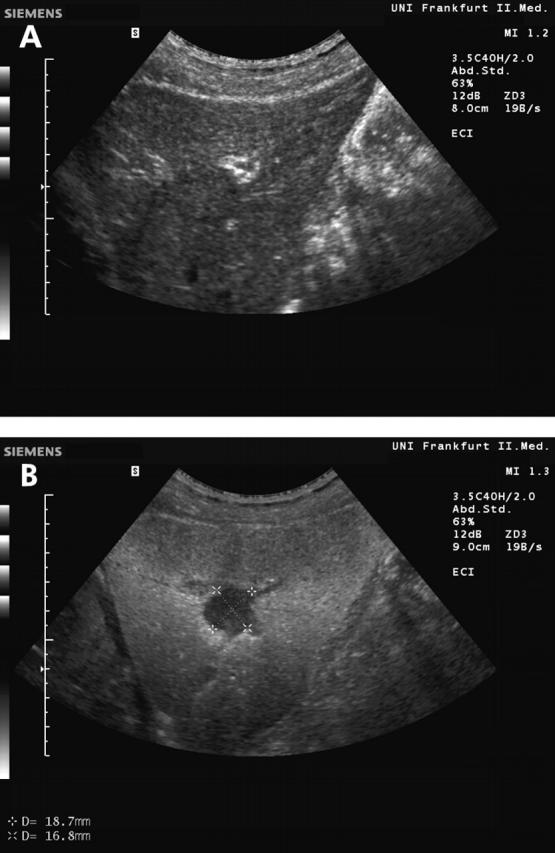
Malignant liver lesion in a patient with colorectal carcinoma and histologically proven liver metastasis. The unenhanced baseline scan shows a slightly heterogeneous liver parenchyma and one focal lesion in the left lobe of the liver (A). Phase inversion ultrasound in the late phase after contrast administration revealed a typical hypoechoic lesion, indicating malignancy (B).