
Figure 2.
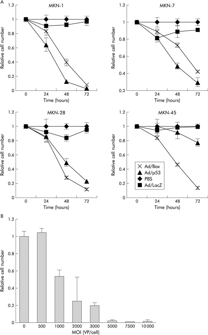
(A) Cell viability, as determined using a colorimetric assay with 2,3-bis-(2-methoxy-4-nitro-5-sulphenyl)-(2H)-tetrazolium-5-carboxanilide (XTT), after infection. The viability value was expressed relative to that of cells infected with phosphate buffered saline (PBS), which was arbitrarily referred to as 1. Values for the Ad/Bax and Ad/p53 groups were significantly different from those for the PBS and Ad/LacZ groups in MKN-1, MKN-7, and MKN-28 whereas only treatment with Ad/Bax significantly differed from that in the other groups in MKN-45 cells. Values are means (SD) of one of two similar quadruplicate studies for each groups. (B) MKN-45 cells were mock infected or infected with increasing multiplicities of infection (MOIs) of Ad/Bax and then subjected to the XTT assay 72 hours after viral infection. Values are mean (SD). VP, viral particles.