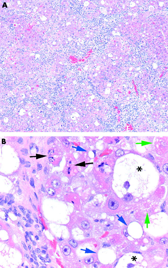
Figure 1.

Morphometric analysis of the cirrhotic liver. After eight weeks of carbon tetrachloride or vehicle treatment, liver tissue was fixed and stained with haematoxylin-eosin. (A) Complete liver cirrhosis with intensive bridging fibrous septum formation is visible. Fibrous septa uniformly affected hepatic tissue, dividing liver parenchyma into small pseudonodules. (B) Liver tissue displayed necroinflammatory damage consisting of inflammatory cell infiltration, both acidophilic necrosis (green arrows) and apoptosis (black arrows), as well as hepatocyte hydropy (*) and fatty degeneration (blue arrows).
