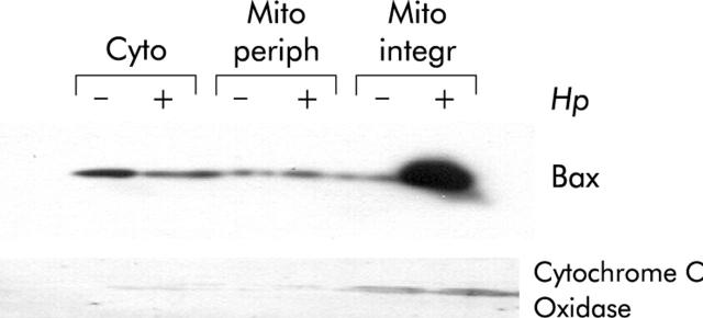
Figure 1.
Helicobacter pylori induced mitochondrial translocation of Bax. AGS cells were cocultured with H pylori for three hours (+) or cultured in medium alone as a control (−). Cytosolic (Cyto), mitochondrial peripheral (Mito periph), and integral (Mito integr) proteins were loaded onto sodium dodecyl sulphate-polyacrylamide gels followed by immunoblotting for Bax and cytochrome c oxidase. Mitochondrial translocation of Bax is shown in samples treated with H pylori. There was a decrease in cytosolic Bax in AGS cells incubated with H pylori and a concomitant increase in mitochondrial membrane integrated Bax. The presence of the control protein, cytochrome c oxidase, in the mitochondrial membrane fractions confirms the quality of the lysates.

