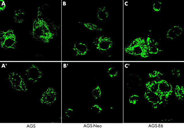
Figure 4.
AGS, AGS-E6, and AGS-neo cells transfected with mito-green fluorescent protein (GFP) were treated with Helicobacter pylori and visualised by confocal microscopy. Mitochondrial morphology was assessed after H pylori treatment. At 10 hours of treatment, more than 70% (5%) of AGS cells showed a fragmented mitochondrial phenotype (A′). Comparable results were obtained in AGS-neo cells (B′). The typical reticulotubular mitochondrial phenotype of healthy AGS cells disintegrated into multiple small rounded organelles (A, A′). In contrast, in AGS-E6 cells with inactivated p53 function, less than 15% (5%) of cells underwent mitochondrial fragmentation under identical experimental conditions (C, C′).

