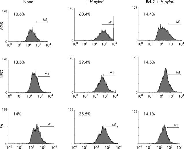
Figure 7.
Caspase-3 activity was inhibited in gastric cells with forced Bcl-2 expression treated with Helicobacter pylori. AGS, AGS-E6 (E6), or AGS-neo (NEO) cells were cocultured with or without H pylori for four hours. Caspase-3 activity was measured with a peptide substrate for caspase-3 which when cleaved emits green fluorescence (excitation, 505 nm; emission, 530 nm). Data were accumulated by analysing an average population of 20 000 cells by flow cytometry. In parallel experiments, AGS, AGS-E6, or AGS-neo cells transfected with Bcl-2 (to inhibit apoptosis) were stained as explained above. Caspase-3 activity was quantified by flow cytometry analysis of untransfected cells versus Bcl-2 transfected gastric cells after H pylori treatment for the indicated time periods. Bcl-2 transfected cells exhibited significantly reduced caspsase-3 activity four hours after H pylori treatment (p<0.05). In contrast, untransfected cells exhibited much higher caspase-3 activity. Values represent the average of triplicate samples.

