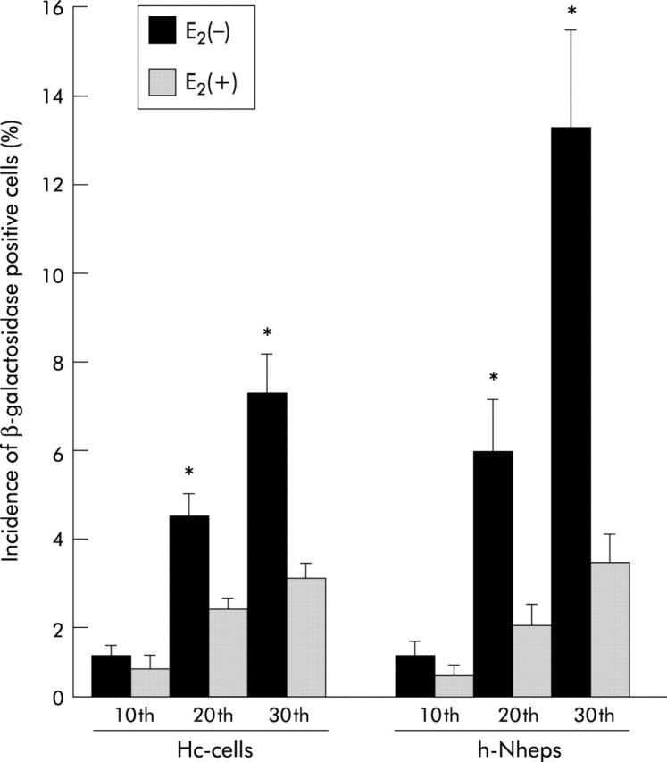
Figure 7.

Incidence of positivity for β-galactosidase in human normal hepatic cells (Hc-cells and h-Nheps). The number of β-galactosidase positive cells increased with accumulated passages, and was significantly decreased in oestradiol (E2) treated cells in comparison with non-treated cells after the same number of passages (*p<0.05). Two researchers microscopically evaluated 500 cells five times, and determined the percentage of those staining positive for β-galactosidase.
