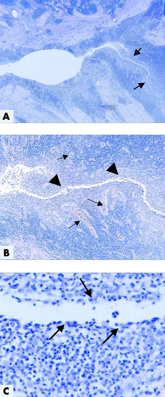
Figure 1.

Histopathological picture of colonic Crohn’s disease with a fistula. (A) At low magnification, the mucosa is destroyed, ulcerated, and replaced by granulation tissue, including a dense capillary network. The surface is covered by fibrin and neutrophils. In the right half, there is a deep fistula which infiltrates the muscularis propria (arrows) (haematoxylin-eosin (H&E), ×16). (B) The central fissure (arrowheads) is branching deep into the underlying tissue, lined by neutrophils, granulation tissue with numerous capillaries (arrows), histiocytes, and lymphocytes (H&E, ×50). (C) At the surface of the central fissure, there is a small layer of histiocytes and neutrophils (arrows). Lining epithelium is not detectable (H&E, ×400).
