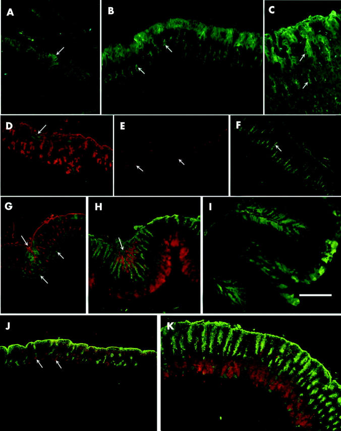
Figure 3.

Immunohistological localisation of trefoil factors (TFFs) during postnatal development of the oxyntic mucosa of normal (A–I) and TFF1 knockout (J, K) mice. Paraffin sections were incubated with rabbit antimouse TFF1 (A–C), TFF2 (D–F), and TFF3 (G–K). Antigen-antibody binding sites were visualised by the fluorescine isothiocyanate (FITC) conjugated goat antirabbit IgG (green: A–D, F, G, I) or with tetramethylrhodamine isothiocyanate (TRITC) conjugated goat antirabbit IgG (red: E, H, J, K). Mucus secreting pit cells were labelled with either TRITC (red: D, G) or FITC (green: H, J, K) conjugated to lectin. (A) P1: folded epithelial lining exhibiting small patches of TFF1 expressing cells (arrow). (B) P3: pit cells lining the luminal surface labelled with anti-TFF1 as some cells in the developing epithelial buds (arrows). (C) P21: localisation of TFF1 in the cells lining the luminal surface and pit region (upper arrow). Some cells in a region corresponding to the isthmus also exhibited labelling in the apical cytoplasm (lower arrow). (D) P1: pit cells (red) were located along the luminal surface and in the primordial buds. Small patches of TFF2 expressing epithelial cells (arrow) were found mainly at the luminal surface. (E) P3: TFF2 expressing cells (arrows) were located in the middle of primordial epithelial buds. (F) P60: stomach shows an increase in mucosal thickness and expansion in the area of cells labelled with TFF2 (arrow) which corresponds to the neck region of the gastric glands. (G) P7: pale TFF3 staining (green) was seen in pit cells (red) along the surface and intensified in pit cells forming a mucosal crease (upper left arrow). Deep in the mucosa there were also several scattered parietal cells expressing TFF3 (lower arrows). (H) P21: in addition to pit cells, TFF3 was expressed in zymogenic cells (red) seen at the bottom of the mucosa. The arrow points to a mucosal crease with many Ulex europaeus agglutinin (UEA) labelled (green) pit cells expressing TFF3 (red). Note that away from the mucosal crease, UEA labelled pit cells do not produce TFF3. (I) P60: TFF3 expressing cells (green) were located in the pits forming the mucosal creases (top and lower left) and in zymogenic cells (centre and right side). (J) P3: pit cells (green) were seen along the surface and in the developing epithelial buds. TFF3 staining (red) was seen in pit cells along the surface and in parietal cells (arrows). (K) P21: pit cells (green) were seen along the surface and in the pits of developing nascent glands. TFF3 staining (red) was seen deep in the mucosa where zymogenic cells are located. Bar = 50 (A–C), 70 (D–K) µm.
