
Figure 5.
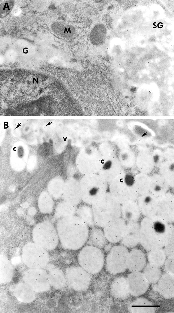
Immunogold localisation of trefoil factors 2 (TFF2) and 3 (TFF3) in normal oxyntic mucosae of adult (postnatal day 60) mice. (A) Supranuclear cytoplasm of a neck cell showing part of the nucleus (N) and the Golgi apparatus (G). Several gold particles were seen on the contents of the Golgi and the newly produced secretory granule (SG), indicating the presence of TFF2. No gold particles were seen on the mitochondria (M). (B) Apical cytoplasm of a pit cell, as seen in the upper segment of the pit. The cytoplasm was packed with many secretory granules and some exhibited a dense core (c). Several gold particles bound to the secretory granule contents. Note that the dense cores exhibited more gold particles than the pale mucinous contents. The apical membrane of the cell projected a few microvilli (v) towards the gland lumen which contained exocytosed secretory material bound to some gold particles (arrows). Bar = 300 nm.