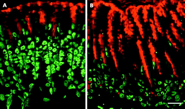
Figure 6.
Immunohistological studies of pit and parietal cell lineages in normal and trefoil factor 1 (TFF1) knockout mice. (A) Normal stomach: note that pit cells (red) were seen along the luminal surface and in the pits which form about one third of the mucosal thickness, whereas parietal cells (green) were scattered in the lower two thirds of the mucosa. (b) TFF1 knockout stomach: pit cells expanded and occupied the upper two thirds of the mucosa, whereas parietal cells were reduced in number and became localised mainly in the lower third of the mucosa. Bar = 30 µm.

