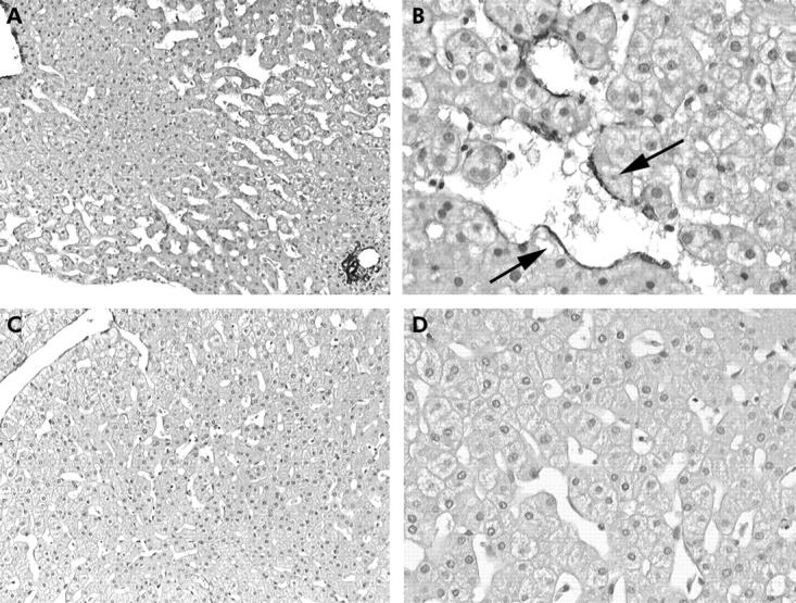
Figure 1.

Marked sinusoidal dilatation, prominent in the mediolobular zone of the lobule (zone 2) (A, ×100) with detection of α-smooth muscle actin positive perisinusoidal cells (B, arrows, ×250). Moderate sinusoidal dilatation in zones 2 and 3 (C, ×100), with no α-smooth muscle actin positive cells detected (D, ×250).
