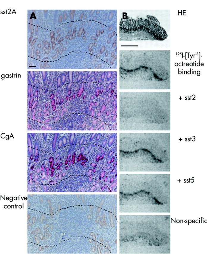
Figure 1.

Comparison of immunohistochemistry (A) and receptor autoradiography (B) in the gastric antrum. (A) Localisation of immunohistochemically detected cells in the gastric antrum. 1st row: somatostatin receptor 2A (sst2A) cells lie at the neck of the glands. They represent a linear zone, indicated between the broken lines. Staining of the more superficial layers was non-membranous and non-specific. 2nd and 3rd rows: Gastrin and CgA cells, respectively, were localised in the same region as sst2A cells. 4th row: Peptide preabsorption as a negative control for sst2A showing residual non-specific non-membranous staining. Bar 100 μm. (B) Receptor autoradiographic illustration of sst receptor subtypes in the gastric antrum. 1st row: Haematoxylin and eosin (HE) stained section. 2nd row: Autoradiogram showing total binding of the ligand 125I-[Tyr3]-octreotide at the neck of the glands. 3rd row: Autoradiogram showing 125I-[Tyr3]-octreotide binding in the presence of 100 nM of an sst2 selective ligand (L-779,976). Complete displacement of the radioligand is seen. 4th and 5th rows: Autoradiograms showing 125I-[Tyr3]-octreotide binding in the presence of 100 nM of an sst3 selective ligand (315-164-15) and 100 nM of an sst5 selective ligand (L-817,818). No displacement of binding was seen. 6th row: Non-specific binding (in the presence of 100 nM octreotide). Complete displacement of labelling by the sst2 selective ligand indicates the presence of sst2 receptors. Bar 1000 μm.
