
Figure 5.
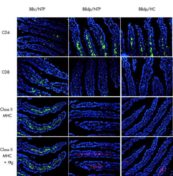
Diet and enteropathy in the BB rat. Immunohistological appearance of the jejunum in 70 day old BB diabetes prone (BBdp) and control (BBc) rats fed hydrolysed casein (HC) and NTP diets. Frozen sections were stained for CD4+, CD8+, and class II MHC+ cells using Alex Fluor 488 (green) and for αE integrin using Alex Fluor 647 (red). Nuclei were stained with DAPI (blue) (×200 magnification).