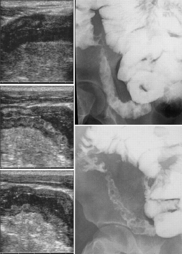
Figure 1.

Oral contrast US (left) and barium enteroclysis (right) in a patient with Crohn’s ileitis. Note at US longitudinal sections (from the top to the bottom) the anechoic contrast flow through the terminal ileum and the distension of bowel walls with a clear definition of the cobblestone mucosal pattern (white arrows) as well as the reduction of BWT from 8.1 mm to 6.2 mm.
