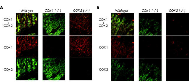
Figure 1.
Cyclooxygenase (COX)-1 and COX-2 immunofluorescence. Sections of fixed mouse corpus were immunolabelled with goat antimouse COX-1 and rabbit antimouse COX-2 polyclonal antisera. Immunofluorescence for COX-1 (red, Alexa 546) and COX-2 (green, Alexa 488) was evaluated in wild-type, COX-1 (−/−), and COX-2 (−/−) mice. Confocal microscopy imaged immunofluorescence at the base of gastric glands (A) and at the gastric surface (B).

