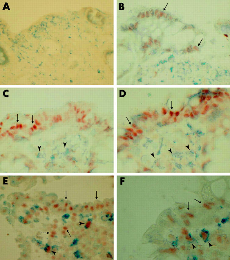
Figure 5.

Double immunohistochemical detection of activated nuclear factor κB (NFκB) and cellular markers for macrophages (CD68) in paraffin embedded formalin fixed biopsies from patients with uninflamed bowel (A, B), collagenous colitis (C, D), or ulcerative colitis (E, F). Negative NFκB staining (A) or weak but distinct focal staining of epithelial cells (B; arrows; red staining) was seen in uninflamed bowel. In collagenous colitis, NFκB nuclear staining was predominantly seen in the epithelium (C, D; arrows) while CD68 positive macrophages (C, D; arrowheads; blue staining) and CD68 negative stromal cells were left unstained. In ulcerative colitis, epithelial cells (E, F; arrows), CD68 positive macrophages (E, F; arrowheads), and CD68 negative stromal cells (E; broken arrows) showed intense nuclear expression of NFκB. Magnification ×100 (A–E) and ×150 (F).
