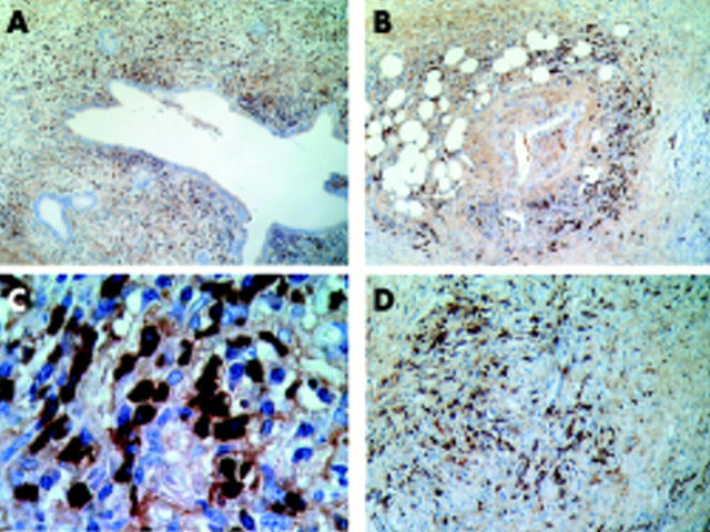
Figure 4.
Representative immunohistochemical images with IgG4 immunostaining. (A) Pancreatic duct surrounded by a severe lymphoplasmacytic infiltrate predominantly of IgG4 positive plasma cells. (B) Prominent IgG4 positive plasma cells within a dense lymphoplamacytic infiltrate surrounding a pancreatic artery. (A) and (B) correspond to case No 1 in table 4 ▶. (C) Abundant IgG4 positive plasma cells together with some IgG4 negative plasma cells, which corresponds to patient No 3 in table 4 ▶. (D) Diffuse fibrosis around a vein with severe mononuclear lymphoplamacytic infiltrate containing abundant IgG4 positive plasma cells, which corresponds to patient No 4 in table 4 ▶. Amplifications were: ×40 for A and B, ×1000 for C, and ×200 for D.

