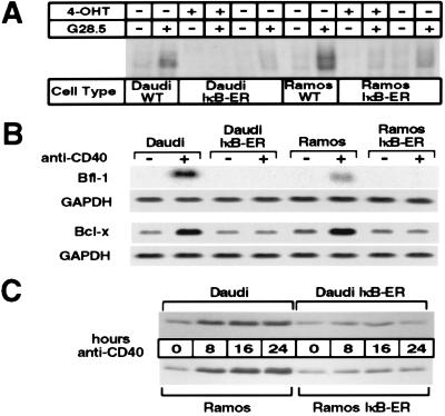
Figure 2.
Cells expressing IκB-ER have a functional block in NF-κB signaling and do not up-regulate Bcl-x and Bfl-1 in response to CD40 activation. (A) Cells were stimulated for 25 min in the presence or absence of 1 μg/ml G28.5 antibody, and the nuclear extracts were then processed as described previously. Six micrograms of nuclear extracts then were used in binding assays with the radiolabeled MHC II oligonucleotide. Cells induced with 4-OHT were pretreated with 200 nM of the synthetic estrogen for 3 hr before G28.5-mediated activation. (B) Daudi, Daudi-IκB-ER, Ramos, and Ramos-IκB-ER cells were pretreated with 4-OHT for 45 min and then cultured for 8 hr in 4-OHT alone or 4-OHT plus G28.5. Eighteen micrograms of total RNA was loaded per lane, and the Northern blots were hybridized with a human Bfl-1 or Bcl-x probe. Northern blots were stripped and glyceraldehyde 3-phosphate dehydrogenase (GAPDH) expression was probed as a standard. (C) Daudi, Daudi-IκB-ER, Ramos, and Ramos-IκB-ER cells were pretreated with 4-OHT for 2 hr and then stimulated with G28.5 in the presence of 4-OHT for different periods of time. Total cell extracts from 1 × 10⁶ cells were loaded per lane, and Western blotting was performed to detect Bcl-x expression.

