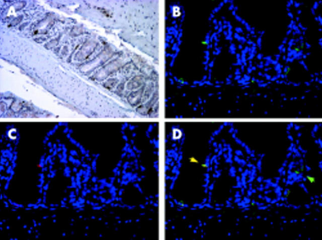
Figure 5.
Immunohistochemical detection of interferon γ (IFN-γ) and asialo-GM1 in the colon of C.B17.SCID mice colonised with Escherichia coli. Colon of SCID E coli mouse stained with (A) IFN-γ antibody followed by DAB development, 20×; (B) asialo-GM1 antibody with Cy2 fluorescent secondary antibody (green), 40×; (C) IFN-γ antibody with Cy3 fluorescent secondary antibody (red), 40×; and (D) colocalisation of asialo-GM1 and IFN-γ with fluorescent secondary antibody, 40×. Yellow arrow = colocalised IFN-γ and asialo-GM1; green arrow = asialo-GM1 only. Immunofluorescent sections (B-D) costained with DAPI to indicate nuclei (blue).

