
Figure 4.
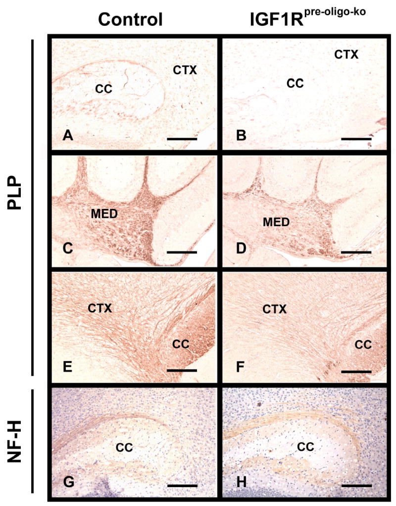
PLP and NF-H immunostaining in brains of IGF1Rpre-oligo-ko mice (panels B, D, F and H) and their littermate controls (panels A, C, E and G). Brains from 2-week-old (panels A, B, C, D, G and H) and 6-week-old (panels E and F) IGF1Rpre-oligo-ko mice and their littermate controls were stained with PLP antibody (panels A, B, C, D, E and F) or NF-H antibody (panels G and H). After NF-H immunostaining, sections were counterstained with hematoxylin. CTX = cerebral cortex, CC = corpus callosum, and MED = cerebellar medulla. The scale bar represents 200 μm in panels A, B, C, D, G and H, and 100 μm in E and F, respectively.