Figure 2.

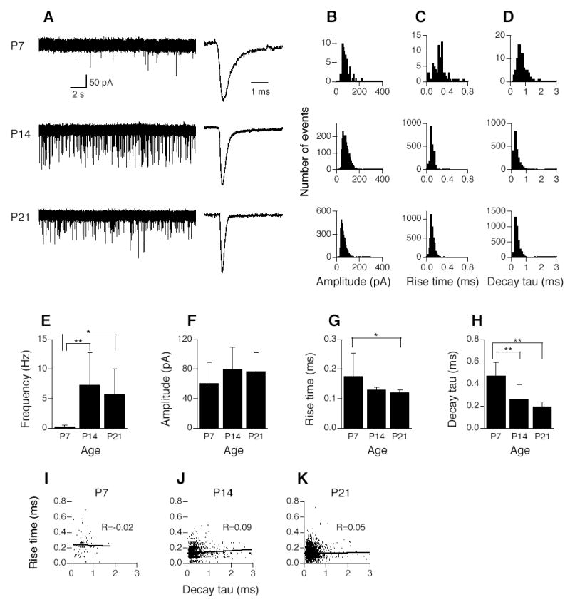
Development of spontaneous miniature EPSCs (mEPSCs) in AVCN bushy cells. A. Shown on the left are three original chart recordings of mEPSCs of AVCN bushy cells from mice ages P7, P14, and P21. Cells were voltage-clamped at −60 mV. Strychnine (1 μM) and gabazine (10 μM) were present in the perfusion solution to block glycine and GABAA receptors. Shown on the right are averaged traces from mEPSCs of 3 individual cells, normalized to their peaks. B–D. Histogram distributions of mEPSC amplitude (bin width of 5 pA), 10–90% rise time (bin width of 20 μs), and decay time constant (decay tau, bin width of 100 μs) of all events of all cells (n=6, 6, 9 cells for P7, P14, and P21, respectively) at the three different ages. Labels indicating age at the far left in A apply to histograms in B–D. E. Frequency of mEPSCs increases significantly from P7 to P14 or P21. No significant difference was detected in mEPSC frequency between P14 and P21. F. Amplitude of mEPSCs shows no difference across ages tested. G. mEPSCs of bushy cells recorded from P21 mice rise significantly faster than those of P7 mice, as evidenced by briefer 10–90% rise time of mEPSCs. The difference between P7 and P14 is not significant (p=0.086). H. Decay tau of mEPSCs gets progressively smaller from P7 to P21. Significant differences were detected by ANOVA (p<0.05), and post hoc Fisher’s tests show that decay tau at P14 is significantly smaller than that at P7, and decay tau at P21 is also significantly smaller than that at P7, but no significant difference was detected between P14 and P21. Note that to generate graphs F, G, and H, an averaged trace was obtained for each cell by averaging all detected events in that single cell. Amplitude, 10–90% rise time, and decay tau of the averaged mEPSCs were measured, then data were pooled in each age group. Means ±standard deviations are shown in E–H. *p<0.05, **p<0.01 (ANOVA post hoc Fisher’s test). I–K. Plots of 10–90% rise time against decay tau of mEPSCs in AVCN bushy cells at ages P7, P14, and P21. No correlations between mEPSC 10–90% rise time and decay tau were found in AVCN bushy cells at the ages tested, indicating a lack of dendritic filtering, consistent with the fact that excitatory inputs from the auditory nerve impinge onto the soma, not the dendrites, of bushy cells.
