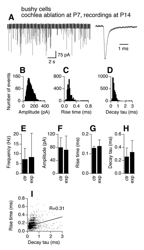
Figure 4.

Effects of cochlea ablation performed during the critical period on mEPSCs of AVCN bushy cells. The frequency and kinetics of mEPSCs in AVCN bushy cells 7 days after unilateral cochlea ablation (surgery performed at P7) are similar to those recorded from normal mice of the same age (P14). A. Original chart recording of mEPSCs of a bushy cell representative of mEPSC frequency of the group, and an averaged trace of all events detected from another cell representative of mEPSC kinetics of the group. The averaged trace was normalized to its peak with the averaged traces in Figure 2A. B–D. Histogram distributions of mEPSC amplitude (bin width of 5 pA), 10–90% rise time (bin width of 20 μs), and decay tau (bin width of 100 μs) of all events from 7 bushy cells obtained from the AVCN ipsilateral to the cochlea ablation. E–H. Unpaired t-tests detected no difference in average frequency, amplitude, 10–90% rise time, and decay tau of mEPSCs between control (same data for P14 in Fig. 2E–H) and the experimental group (p>0.05). Means ±standard deviations are shown. ctr: control; exp: experimental group. I. Seven days after unilateral cochlea ablation (surgery performed at P7), a positive correlation between mEPSC 10–90% rise time and decay tau is seen in bushy cells in the AVCN ipsilateral to the cochlea ablation, suggestive of emergence of dendritic filtering in bushy neurons that survived the surgery.
