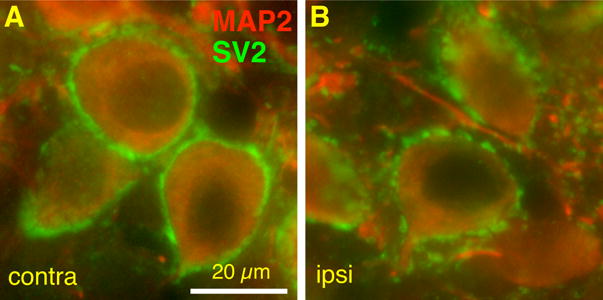
Figure 8.

Effects of cochlea ablation performed after the critical period on SV2 expression in AVCN bushy cells. Representative sections showing SV2 (green) and MAP2 (red) labeling of bushy cells contralateral (contra, A) and ipsilateral (ipsi, B) to the cochlea ablation. Note the presence of the ring of SV2 labeling around the somata in both panels. However, the SV2 label on the ipsilateral somas appears less continuous than on the contralateral side. Scale bar, 20 μm.
