Figure 3.
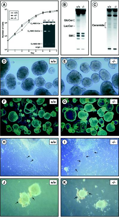
In vitro growth and differentiation of ES cells in the absence of GSL synthesis. (A Left) Growth of wild-type (+/+), UgcgΔEX7Neo/+ (+/−), and UgcgΔEX7Neo/ΔEX7Hygro (−/−) ES cells. ES cells (5 × 10⁴) were seeded into 60-mm plates containing mitomycin C-treated embryonic fibroblasts to initiate the experiment. Each day the medium was changed and the cells from one plate were harvested and were counted. (Inset) Glucosylceramide synthase activity in wild-type (+/+), UgcgΔEX7Neo/+ (+/−), and UgcgΔEX7Neo/ΔEX7Hygro (−/−) ES cells. Each cell extract, corresponding to 50 μg of protein, was incubated with C6-NBD-ceramide and UDP-glucose. After incubation, lipids were separated by high-performance thin-layer chromatography (HPTLC) and were visualized by UV illumination. The positions of the C6-NBD derivatives of glucosylceramide (C6-NBD-GlcCer) lactosylceramide (C6-NBD-LacCer) and sphingomyelin (C6-NBD-SM) are indicated (B) HPTLC analysis of neutral glycolipids isolated from wild-type (+/+) and UgcgΔEX7Neo/ΔEX7Hygro (−/−) ES cells. The positions of glucosylceramide (GlcCer), lactosylceramide (LacCer), and sphingomyelin (SM) are indicated. (C) HPTLC analysis of ceramide levels in wild-type (+/+) and UgcgΔEX7Neo/ΔEX7Hygro (−/−) ES cells. The position of ceramide is indicated. (D and E) Wild-type (+/+) and UgcgΔEX7Neo/ΔEX7Hygro (−/−) embryoid bodies after 3 days of culture (×60). (F and G) Wild-type (+/+) and UgcgΔEX7Neo/ΔEX7Hygro (−/−) cystic embryoid bodies after 22 days of culture (×20). (H and I) Retinoic acid-induced neuronal differentiation of wild-type (+/+) and UgcgΔEX7Neo/ΔEX7Hygro (−/−) ES cells (×100). Arrows indicate neurite formation. (J and K) Erythropoietin-induced hematopoietic differentiation of wild-type (+/+) and UgcgΔEX7Neo/ΔEX7Hygro (−/−) ES cells (×100). Arrows indicate clusters of red erythroid cells surrounding embryoid bodies.

