Figure 4.
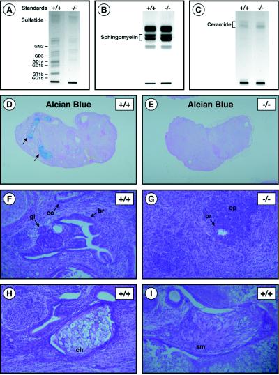
In vivo differentiation of teratomas in the absence of GSL synthesis. (A) Acidic lipid fractions from of wild-type (+/+) and UgcgΔEX7Neo/ΔEX7Hygro (−/−) teratomas analyzed by HPTLC. The position of GSL standards are indicated on the left. (B) HPTLC analysis of sphingomyelin levels from of wild-type (+/+) and UgcgΔEX7Neo/ΔEX7Hygro (−/−) teratomas. The position of sphingomyelin is indicated. (C) HPTLC analysis of ceramide levels in wild-type (+/+) and UgcgΔEX7Neo/ΔEX7Hygro (−/−) teratomas. The position of ceramide is indicated. (D and E) Sections of wild-type (+/+) and UgcgΔEX7Neo/ΔEX7Hygro (−/−) teratomas stained with Alcian blue (×5). Arrows indicate Alcian blue-positive cartilage in the wild-type tumor but not found in the mutant tumor. (F) H & E-stained section of the wild-type teratoma showing well differentiated epithelial tissues (×100). gl, glands; br, bronchial epithelium; co, colonic epithelium. (G) H & E-stained section of UgcgΔEX7Neo/ΔEX7Hydro (−/−) teratoma showing poorly differentiated epithelial tissue (ep) and small focus of bronchial epithelium (br) (×100). (H) H & E-stained section of a wild-type teratoma showing chondrocytes (ch) (×100). (I) H & E-stained section of the wild-type teratoma showing smooth muscle (sm) (×100).

