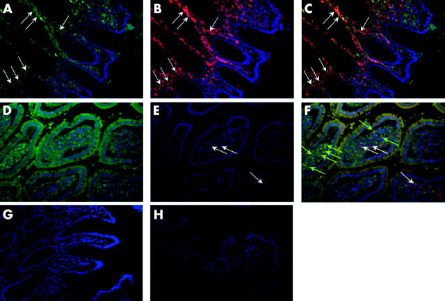
Figure 7.
Immunofluorescence for glycoprotein 96 (gp96) expression in the intestinal mucosa of patients with no inflammation and with Crohn’s disease (CD). Paraffin embedded sections were cut and gp96 was detected with a monoclonal antibody. In a second step, a secondary Alexa Fluor 594 conjugated goat antirat IgG antibody was used (red). CD68 was also detected with a monoclonal antibody and in a second step an Alexa Fluor 488 chicken antimouse IgG (H+L) was used (green). Counterstain was done with 4′,6-diamidino-2-phenylindole (blue). In non-inflamed mucosa (A–C), most of the intestinal macrophages (A, green, arrows) were gp96 positive (B, red; C, merge, arrows). Only very few intestinal macrophages (D, green) from CD patients were gp96 positive (E, red; F, merge, white arrows). No fluorescence was detected using rat IgG2a or mouse IgG1 isotype control (G, H). Original magnification ×200.

