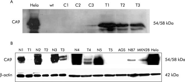
Figure 1.
(A) Western blot analysis after carbonic anhydrase IX (Ca9) transfection of AGS cells. Untreated Hela cells served as a positive control (Hela). Wild-type (wt) AGS cells exhibited no Ca9 expression. Transfection of AGS cells with the empty vector resulted in no detectable Ca9 protein levels after one and two days (C1, C2) After three days, low Ca9 expression was noted which may have been induced by intercellular contact and thus was cell density dependent (C3). AGS cells transfected with Ca9 cDNA exhibited significant Ca9 protein levels after one, two, and three days (T1–T3). (B) Western blot analysis also revealed reduced Ca9 protein levels in gastric cancer (T) compared with non-neoplastic gastric mucosa (N). Ca9 was identified as a protein of 54 and 58 kDa. β-Actin protein levels were assessed for standardisation of protein levels. No Ca9 protein was detected in AGS cells whereas low levels were found in N87 and MKN28 cells. Hela cells served as a control.

