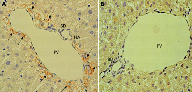
Figure 2.
Immunohistochemical analysis of liver tyrosine hydroxylase (TH) positive nerve fibres showing the region of the portal triad in the liver two weeks after the operative procedures. TH positive nerve fibres (arrows) are seen around the portal vein (PV), bile duct (BD), and hepatic artery (HA) in sham operated mice (A) but not in mixed sympathectomised animals (B). Magnification ×400. The results are representative of three sets of experiments performed on two animals each.

