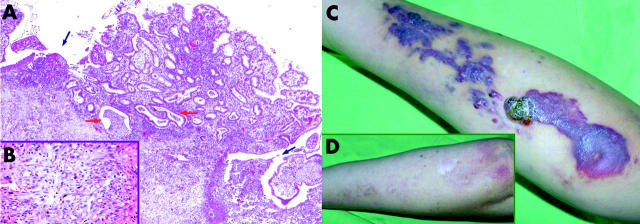
Figure 1.
(A) Histological signs of ulcerative colitis and Kaposi’s sarcoma (KS) from the resected colon. Ulcers (blue arrows) at the base of a pseudopolyp and crypt abscesses (red arrows) can be seen (haematoxylin-eosin staining, 100× magnification). (B) Typical features of KS can be identified in the submuscular connective tissue layer of the colon (haematoxylin-eosin staining, 400× magnification). (C) Kaposi’s sarcoma on the forearm of the patient, and the same region a year after operation (D).

