Figure 3.

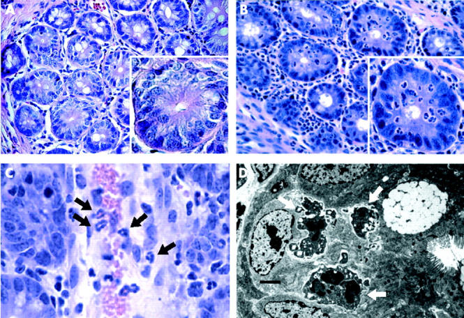
Microscopic features of neutrophil infiltrates in caecum of FABP-rtTA × TRE2IL-8 mice after DOX induction. (A) (B) H&E stained sections of caecum from FABP-rtTA × TRE2IL-8 mice were prepared in uninduced mice (A) and 48 hours following DOX induction (B). The main images are at 200× magnification and the single crypts in the inset at 600×. A dense infiltrate of neutrophils is observed in the lamina propria and intestinal epithelium following DOX induction, but neutrophils did not migrate into the crypt lumen and epithelial cellular damage was not observed. (C) Neutrophils (black arrows) are undergoing diadepedesis through postcapillary venules in the lamina propria after DOX induction (600×). (D) Transmission electron microscopy images in DOX-induced FABP-rtTA × TRE2IL8 mice show neutrophils with intact granules (white arrows) in the paracellular space (scale bar = 1 μm).
