
Figure 3.
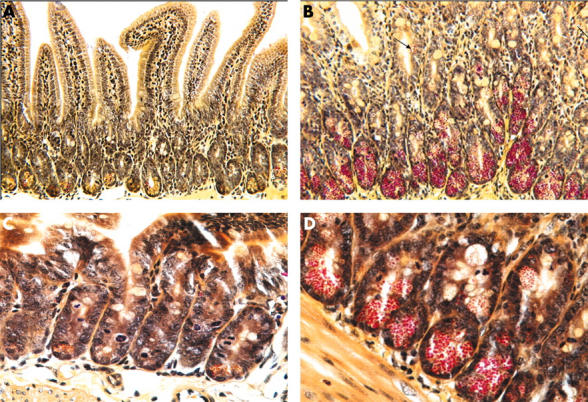
Low (A, B) and high (C, D) power views of phloxine-tartrazine stained sections of uninfected control (A, C) and T spiralis infected (B, D) murine small intestine. A few Paneth cells (with predominantly yellow granules) are present at the base of uninfected crypts. In T spiralis infected intestine, there is a marked increase in the number of Paneth cells (with red granules). Moreover, intermediate cells (arrowed) are seen in some villi. Two (A, B) of the figures are reproduced from Kamal and colleagues,28 with permission from Blackwell Publishing Ltd.