
Figure 4.
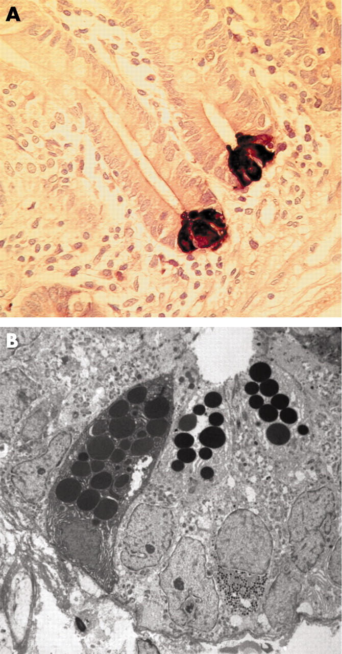
(A) Section of human jejunal mucosa showing human defensin 5 immunoreactive Paneth cells at the base of crypts. (B) Transmission electron micrograph of human jejunal crypt showing three Paneth cells with electron dense granules in the apical cytoplasm.