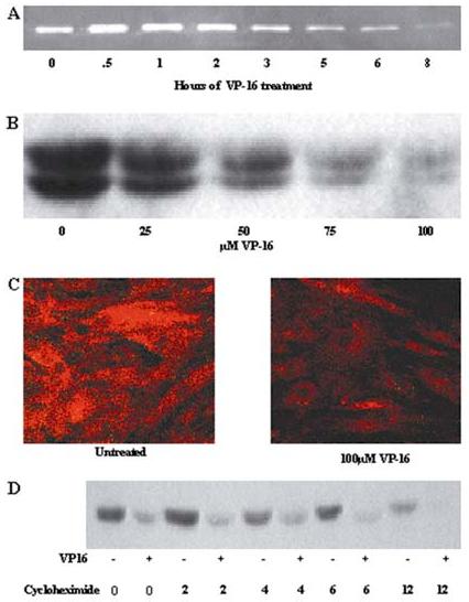
Figure 1.

MMP-2 protein is dysregulated in BMSCs exposed to VP-16. (A) BMSC supernatants were conditioned in serum-free medium for 8 hours. At each time point (30 minutes OE 8 hours) 100:M VP-16 was added to the conditioned BMSCs media. After 8 hours the supernatants were collected and gelatin zymography performed to detect MMP-2. (B) Supernatants were collected from VP-16 treated BMSCs and concentrated as described in Materials and Methods. MMP-2 monoclonal antibody was used to detect MMP-2 protein in VP-16 treated groups compared to supernatants collected from untreated control BMSCs. Data are representative of three independent experiments. (C) BMSCs treated with 100μM VP-16 for 24 hours were fixed and stained with MMP-2 monoclonal antibody and subsequently incubated with PE-tagged secondary antibody. Fluorescence was detected by confocal microscopy. (D) Confluent BMSCs were treated with either VP-16 or VP-16 and cycloheximide for 2-24 hours. At each time point the supernatants were collected, concentrated 10X, and subjected to western blot with antibody specific to MMP-2.
